
Some injuries hide. An athlete has clear pain in a defined spot; the clinical examination points to bone, yet the X-ray looks normal, and even the MRI is inconclusive. This is exactly the situation a SPECT CT bone scan is built for. It is one of the most useful problem-solving scans we have when the question is not “is there a structural abnormality?” but “which of these abnormalities is actually generating the pain?”
A SPECT CT bone scan combines two different kinds of information in a single study: a map of where bone is active and remodelling, and a detailed anatomical picture of the bone itself. Fusing the two lets us place a hot spot of bone turnover precisely onto the structure that produces it. For example, a stress fracture, a cracked pars, or an irritable facet joint may all be causing pain. On this page, we explain what the scan is, why it answers questions other scans cannot, and the specific problems we use it for in sport.
What is a SPECT CT bone scan?
The name describes three parts working together. A conventional bone scan uses a small dose of a tracer (a technetium-99m bisphosphonate) that travels through the bloodstream. It attaches to active bone areas. Anywhere the skeleton is repairing, remodelling, or reacting to injury lights up as a “hot spot.” A bone scan’s strength is its sensitivity: it can detect changes in bone biology long before they show up as a crack or a line on an X-ray.
Its weakness is the flip side of that strength. Many different things increase bone turnover — a fresh fracture, old arthritis, a healing bruise, a degenerative disc. On a simple bone scan, they can all look like a similar area of increased activity. The picture also lacks depth, just as a single front-on X-ray cannot tell you how deep an object sits.
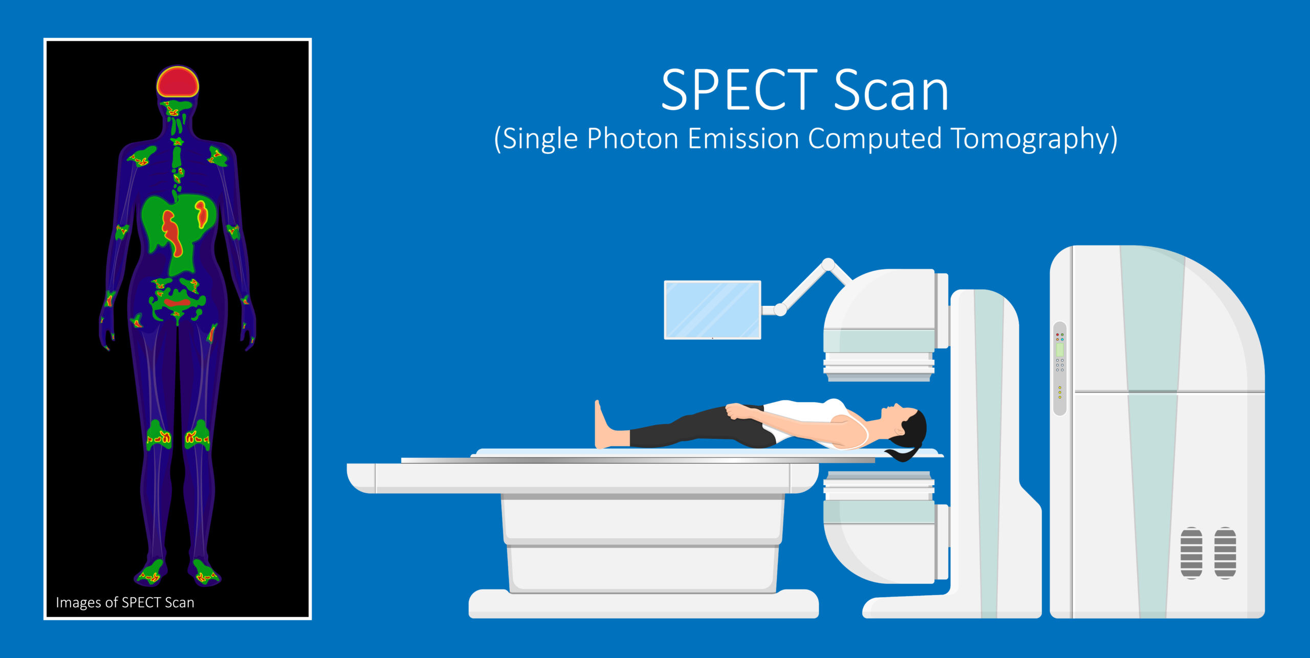
SPECT (single-photon emission computed tomography) solves the depth problem by rotating the camera around you and reconstructing the tracer signal in three dimensions. CT (computed tomography), performed on the same machine in the same sitting, adds anatomy. Modern software then fuses the two datasets so the functional hot spot from the SPECT sits exactly on top of the anatomy from the CT. The result is a scan that shows both that a piece of bone is irritated and which piece it is. Studies consistently show that this fusion improves diagnostic accuracy.

Why we use it: finding the pain generator
Most sports imaging starts with X-ray, ultrasound, or MRI, and for most problems that is exactly right. MRI is particularly good for soft tissues such as muscles, tendons, ligaments, and cartilage. It also uses no radiation. We are not trying to replace it.
However, sometimes the scans show several possible abnormalities and none of them clearly explains the symptoms. A middle-aged runner’s lumbar MRI might show wear at three different levels. A footballer’s foot might show mild arthritis at a joint and tendon swelling. MRI is very good at showing what a structure looks like, but it can’t always tell you which finding is painful.
A SPECT CT bone scan answers that question precisely. The tracer concentrates where bone is genuinely painful, so when we fuse it with the anatomy we can say, with real confidence, “this joint is the active one,” or “this is true bone stress”. Often, it provides a clear target, enabling precise treatment such as a guided injection or rehabilitation.
When a SPECT CT bone scan is useful in sport
Bone stress injuries and stress fractures
Bone stress injuries sit on a spectrum, from early stress reaction through to a frank stress fracture. X-ray is often inaccurate, especially early on. MRI can also miss stress fractures, particularly in certain areas such as the anterior shin. A SPECT CT bone scan detects the increased bone turnover of a stress injury early and then localises it to the exact bone. This matters enormously in sport, because some stress fractures are low-risk and heal with simple rest. However, others are high-risk and need careful protection. Knowing precisely where the injury sits — and confirming it is biologically active — guides how aggressively we need to offload it. It is also valuable when pain persists, and you need to know whether an old injury has truly healed or is still active.
Pars defects and spondylolysis (back pain in young athletes)
Low back pain in a young athlete who does a lot of extension and rotation — gymnasts, cricketers (especially fast bowlers), dancers, rowers, footballers — always raises the question of a pars stress injury. This is a stress fracture of a small bony bridge in the back of the vertebra. We often use MRI to screen suspected cases. However, sometimes, MRI scans miss these injuries. This is a classic indication for SPECT CT: young adolescent athletes with ongoing back pain and a normal MRI scan. SPECT localises the injury to the back of the vertebra, and CT then shows whether a pars defect is present and whether its edges look acute or old. That distinction directly changes management. It often decides whether a young athlete needs to stop and protect the spine or can be managed differently.
Low back pain and the facet joints
In adults, chronic mechanical low back pain is common, and the MRI frequently shows degenerative change at several levels. Sometimes, it is essential to know which level is causing the pain. A SPECT CT bone scan is a well-recognised tool here because tracer uptake indicates which area is causing the pain. This is particularly helpful when considering treatment such as a facet joint injection, because it directs the needle to the joint most likely to respond.
Sacroiliac joint pain
Pain over the back of the pelvis and buttock is notoriously hard to pin down, and the sacroiliac joint is one of several possible sources. A SPECT CT bone scan can show whether the sacroiliac joint is genuinely more active, helping distinguish an active joint from normal changes seen on other imaging. It helps decide whether a targeted injection is worthwhile.
Hip and groin pain
The hip is a frequent source of diagnostic uncertainty in athletes. A SPECT CT bone scan can help clarify unexplained hip and groin pain when other imaging is equivocal. For example, stress fractures around the hip and pelvis that are difficult to see on plain films or MRI. It can also help assess conditions such as early avascular necrosis and transient osteoporosis of the hip when the clinical picture is unclear.
Foot and ankle
The foot and ankle have many small bones and joints packed into a tiny space, making it genuinely hard to identify which structure is causing the pain. SPECT CT is particularly valuable here. It helps localise stress fractures, pinpoint an active osteochondral (cartilage-and-bone) lesion in the ankle, and identify painful accessory bones. By showing which is biologically active, the scan tells us what to treat.
Assessing pain after bony surgery
When pain continues, or returns after surgery involving metalwork — spinal fusion, or hardware in a limb — MRI is often degraded by metal artefact, and CT alone cannot always tell whether bone has healed. Because metal doesn’t block the functional signal of a bone scan the same way, a SPECT CT bone scan can provide more information.
How a SPECT CT bone scan is done
The study is straightforward and takes place in two visits on the same day.
First, a doctor gives a small injection of the radioactive tracer into a vein in your arm. For many bone questions, we take images a few hours later to give the tracer time to be taken up by the skeleton and cleared from the rest of the body. So there is usually a waiting period of around two to four hours between the injection and the main scan. For some questions, particularly where infection or inflammation is a consideration, early images of blood flow are taken soon after the injection as well (a “three-phase” study).
When it is time for the scan, you lie still on a bed while the camera slowly rotates around the area of interest to capture the SPECT images. The CT is acquired on the same machine in the same position. The scanner is open, not an enclosed tunnel like an MRI. No additional preparation is needed beyond what the department advises. You can eat and drink normally unless told otherwise.
Afterwards, you can go straight back to your normal day. The tracer decays and is cleared naturally, and drinking water helps flush it through.
SPECT CT bone scan versus MRI and an ordinary bone scan
It helps to see where each test fits rather than thinking of one as simply “better.”
An MRI is our first choice for most soft-tissue problems and for many bone problems too. It shows muscles, tendons, ligaments, cartilage, and bone marrow in great detail, and it uses no radiation. For many sports injuries, it is all you need.
A plain (planar) bone scan is highly sensitive to changes in bone biology but lacks the depth and anatomical detail to say exactly what and where a hot spot is.
A SPECT CT bone scan solves that problem. We reach for it when the clinical question is specifically about which bone or joint is the active pain generator. We use it when MRI is uncertain or shows too many possibilities, when metal makes MRI difficult to interpret, or when MRI cannot be performed. In short, MRI tends to answer “what does the structure look like?” while SPECT CT answers “which structure is actually reacting?” The two are complementary, and the skill is in choosing the right one for the right question.
Radiation and safety
A SPECT CT bone scan involves a small amount of radiation from both the tracer and the CT. So, we recommend the scan only when it will change your diagnosis or treatment. The tracer dose is small, allergic reactions are rare, and the tracer clears from your body naturally over hours and days. If you are pregnant, might be pregnant, or are breastfeeding, it is important to tell the clinic beforehand, as this affects whether and how the scan is done. For most adult athletes, the dose is a reasonable trade-off for the precise answer the scan provides.
Frequently asked questions
Is a SPECT CT bone scan the same as an ordinary bone scan?
No, though it builds on one. An ordinary (planar) bone scan produces flat images that are very sensitive but cannot always tell you exactly which structure is involved. A SPECT CT bone scan adds a three-dimensional functional scan and a detailed CT in the same sitting. It then fuses them to map the area of increased bone activity to the responsible bone or joint. It is the same tracer injection, with far more useful images.
How long does a SPECT CT bone scan take?
Generally, it takes several hours in total, but you aren’t scanning the whole time. After the tracer injection, you usually wait about two to four hours while the skeleton takes up the tracer. The scan typically takes about 30 to 60 minutes.
Does a SPECT CT bone scan hurt?
No. The only discomfort is the small needle used to inject the tracer, the same as a blood test. The scan itself is painless — you lie still while the camera rotates around you. The scanner is open, not an enclosed tunnel like an MRI.
Is the radiation dangerous?
The scan uses a small dose of radiation from the tracer and the low-dose CT. The dose is modest. But we recommend the scan only when the results will genuinely change your diagnosis or treatment. The tracer clears from your body naturally over the following hours and days.
Why has my doctor recommended a SPECT CT bone scan instead of an MRI?
MRI is excellent and remains our first choice for most problems, but it answers a slightly different question — what a structure looks like. We choose a SPECT CT bone scan when the key question is which structure is actually active and causing pain. Sometimes we use it when an MRI shows several possible causes without a clear answer, when metalwork from previous surgery makes MRI hard to interpret, or when MRI cannot be performed. It is a problem-solving tool, not a routine first scan.
What happens after the scan?
You can return to normal activity straight away. We review and report the images, then go through the findings with you and fit them to your examination and history. The point of the scan is to sharpen the plan. For example, it can confirm a stress injury so we can protect it properly, or identify the single active joint so we can aim a guided injection precisely.
Final word from Sport Doctor London
The real value of a SPECT CT bone scan is not that it is more powerful than everything else. It is that it answers a very specific question better than many other tests: which of several possible abnormalities is actually causing the pain. Used at the right moment, it turns a confusing picture into a clear target and lets us treat the problem precisely rather than guessing. Used at the wrong moment, it adds radiation and cost without benefit. That judgement — knowing when the scan will change the plan and when it will not — is exactly what a sports and exercise medicine specialist is for.
If you have ongoing bone or joint pain that previous scans haven’t explained, or a stress injury that isn’t settling, we can review your imaging, examine you properly, and decide whether a SPECT CT bone scan is the right next step. Call us on +44 (0) 203 488 0350 or book an appointment to discuss your case with Dr Lorenzo Masci.
Leave A Comment