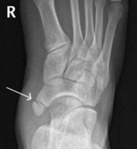
Pain inside the ankle may be due to tibialis posterior tendonitis, mid-foot arthritis, or navicular stress fracture. However, one less common cause of pain is an accessory navicular syndrome or os naviculare. So, what is accessory navicular syndrome, and how do we manage it?
What is an accessory navicular?
The navicular is a wedge-shaped bone connecting the ankle to the mid-foot. The bone has two ossification centres during childhood – one in the centre and one on the inside. In most people, these two centres join together, forming one bone. However, in about 10% of the population, these two centres don’t fuse, meaning there are two bones. The accessory navicular, also called os naviculare, is this extra bone outside the prominent navicular bone. They are joined together by cartilage.
Generally, most people with an os naviculare don’t know they have one. However, sometimes, after an ankle sprain or secondary to chronic rubbing from a shoe, pain occurs inside the ankle. We think pain occurs because the cartilage between the two bones is disrupted.
Accessory navicular syndrome symptoms
Generally, most people with accessory navicular syndrome report the following symptoms:
- Pain on the inside of the ankle or mid-foot
- Swelling or inflammation next to the accessory bone
- A visible prominence or lump corresponding to the accessory navicular
Diagnosis is based on taking a history and examination. Usually, there is tenderness at the accessory navicular bone. In addition, some people with accessory bone have flat feet that pull on the accessory navicular, causing more pain.

Confirming the diagnosis with imaging and ruling out other causes is essential. An X-ray of the ankle will show an extra bone on the inside of the navicular. Also, an MRI scan shows swelling around the bones, suggesting disruption of the cartilage between the bones. Finally, ultrasound demonstrates increased blood flow between the two bones and the involvement of the closely attached tibialis posterior tendon.
Accessory navicular syndrome treatment options
Treatment aims to settle the inflammation. Generally, we start with simple remedies such as regular ice and oral anti-inflammatories such as ibuprofen. Also, custom-made orthotics are adequate to support the medial arch, reducing pressure on the navicular. Finally, an ultrasound-guided cortisone injection into the cartilage between the two bones can sometimes help.
Best shoes for accessory navicular
Generally, we suggest shoes with a wide midsole and a supportive medial arch. Usually, New Balance shoes are well suited to people with this condition.
Os naviculare surgery pros and cons
If conservative measures fail, then surgery can help. For example, if the accessory navicular bone is small, we remove the bone. However, removing the bone and reattaching the tibialis tendon may be needed if the bone is more prominent. In addition, a screw fixing the accessory bone to the prominent bone is sometimes required. Usually, you need a period in a cast and a walking boot after surgery.
More about a cortisone shot for accessory navicular syndrome
Sometimes, we perform injections to reduce pain in Os Naviculare. Generally, we use ultrasound to direct the needle to the exact spot between the two bones. This accuracy is vital to improve the effectiveness and reduce the side effects of cortisone injection, such as skin thinning and tendon rupture.
Final word from Sportdoctorlondon about the accessory navicular syndrome
An accessory navicular is present in about 10% of the population, but only a small number develop pain. Ususally we usually start with simple treatments such as anti-inflammatory treatments and custom-made orthotics first, followed by an injection.
Related conditions:


I’ve found this article to be the most informative of all the articles I’ve read on the navicular bone. The photos are most helpful especially the X-ray. Thank you.
Thanks, Phyllis for the comment.
I’m having accessory navicular bone with inflammation in posterior tibial.. I’m 2.5 months treatment.. can we cure in conservative treatment…
yes I’d always trial conservative management for most conditions including accessory navicular syndrome.
LM
I am suffering from pain due to os navicular for 2 years
Should I go for surgery
Hi Vidya, if you tried other treatments (as outlines in my blog), then surgery would be a reasonable option for persistent pain.
Lorenzo