A pinched nerve in the calf can cause pain in the lower leg or foot — especially when imaging is normal. Nerve pain in the calf is easily missed because it overlaps with muscle and tendon problems. How do we diagnose a pinched nerve in the lower leg, and how do we treat it? A pinched nerve is one of several causes of shin and calf pain, and these nerves form part of the wider group of compressive neuropathies in sport.
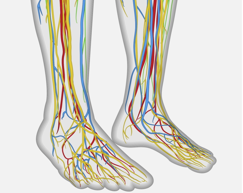
Common pinched nerves in the calf
Common peroneal nerve entrapment
The common peroneal nerve is most often compressed around the fibular neck, where it’s superficial. Athletes feel pain in the outer knee radiating into the outer calf, sometimes with numbness, tingling, or weakness. Pain comes on with running and settles quickly upon stopping. Ankle sprains can tether the nerve near the lateral aspect of the knee.
Superficial peroneal nerve entrapment

The superficial peroneal nerve pierces the deep fascia halfway down the lower leg, and pinching occurs there. Symptoms are a running pain in the outer lower leg and the top of the foot, sometimes with numbness and tingling, which switch off almost immediately when stopped. Ankle sprains or direct trauma are common causes.
Tarsal tunnel syndrome
The tarsal tunnel lies between the tibia and the inner Achilles, carrying the posterior tibial nerve. Compression causes burning or shooting pain in the inner ankle and arch, sometimes with numbness. Causes include a ganglion cyst, arthritic spurs, extra muscles, or ankle twisting. Trapping of a branch of the nerve near the heel causes Baxter’s nerve entrapment, often mistaken for plantar fasciitis.

Sural nerve entrapment
The sural nerve runs down the outer calf alongside the Achilles tendon to the outer foot. Entrapment causes pain along the outer calf into the outer ankle and foot, sometimes accompanied by numbness. It can be trapped where it pierces the deep fascia, near an inflamed Achilles, or by scar tissue after Achilles repair.
Medial plantar nerve entrapment (jogger’s foot)
The medial plantar nerve can be trapped in the inner arch at the Knot of Henry, causing pinpoint pain in the inner arch. Known as a jogger’s foot, it’s often misdiagnosed as plantar fasciitis.
How do we diagnose a pinched nerve in the calf?
It’s challenging because symptoms overlap with other conditions. A key clue: suspect a trapped nerve when imaging, such as an MRI, is normal. The site of symptoms points to the nerve, and nerve pain differs from tendon or joint pain — it’s switched on by walking or running and off by rest. Tapping the nerve at the trapping site (Tinel’s sign) may reproduce the symptoms.
Tests to confirm the diagnosis include:
- Nerve conduction studies — though these can be normal in entrapment
- MRI scan
- Blood tests to exclude nerve dysfunction from diabetes, thyroid disease, or autoimmune disorders
- Ultrasound — increasingly used to track the nerve and find entrapment points, and to guide a local anaesthetic or cortisone injection. Improvement after a cortisone injection makes entrapment more likely.
It’s essential to exclude overlapping conditions — an inflamed nerve from medical disease, local muscle or tendon inflammation, and nerve trapping from above (sciatica or piriformis syndrome).
Does tarsal tunnel syndrome show on MRI?
Sometimes, if a growth is compressing the nerve, an MRI can show it. But MRI doesn’t detect every case of tarsal tunnel syndrome.
How do we treat nerve pain in the lower leg?
Once the diagnosis is confirmed, simple treatments start first:
- Ibuprofen cream or tablets to reduce nerve inflammation
- Physiotherapy — soft-tissue massage, acupuncture, and neural glides to loosen the nerve
- Nerve pain medication such as amitriptyline or duloxetine
- Ultrasound-guided cortisone injection at the site, to confirm and treat
- Nerve hydrodissection, where scar tissue tethers the nerve — floating it free under ultrasound, a technique Dr Masci teaches across the UK and Europe
- Surgery to release the nerve, only after other treatments fail and imaging or injection confirms the cause
Frequently asked questions about a pinched nerve in the calf
Can a pinched nerve in the calf cause numbness in the foot?
Yes. Because these nerves travel to the foot, entrapment in the calf or lower leg often causes numbness or tingling in the foot or ankle, alongside the pain — the pattern depends on which nerve is affected.
Why are my scans normal,l but my calf still hurts?
Nerve entrapment frequently shows normal X-rays and MRIs — a normal scan with ongoing nerve-type pain actually supports the diagnosis. Ultrasound, nerve conduction studies, and a diagnostic injection help confirm it.
Could my calf nerve pain be coming from my back?
Yes. Nerve pain in the lower leg can be referred from the lumbar spine (sciatica) or the buttock (piriformis syndrome) rather than a local calf entrapment. Working out the true source is the key step before treatment, because it changes the plan entirely.
Final word from Sport Doctor London about a pinched nerve in the calf
Unexplained calf, foot, or ankle pain may be a pinched nerve — especially when scans are normal. Diagnosis combines a careful assessment with ultrasound, nerve conduction studies, and sometimes an injection. See a doctor experienced in these uncommon causes of unexplained pain.
To book a one-stop assessment with Dr Masci in London, contact the team here or call +44 (0) 203 488 0350.
I had sciatica for 6 weeks now the pain in back is slowly going .but now the pain is on top of my calf muscle leading to my ankle .many years ago I was involved in a I.e.d explosion in Iraq witch left 3 toes on my right foot with no nerves what can I do to stop the pain at the back of my right calf
Physical therapy is important. I would speak to your doctor about other options: medications such as amitriptyline or possible injections depending on whether your pain is back-related (sciatica) or local.
https://sportdoctorlondon.com/amitriptyline-for-pain-side-effects/
Can a pinched nerve in the calf happen after a dislocation of the knee? He wouldn’t go to a doctor after falling and the knee popped back in the next day while getting him into the shower. Swelling went down significantly. He is using crutches to get around when necessary and has been resting for 3 days(kind of) but now his calf hurts more than the injured knee he says. I haven’t been able to get a look to check for redness or swelling but so far no fever or anything….when do I need to force him to see the doctor?
Hi Aspen, It could be a pinched or damaged nerve after a knee dislocation. You need to see an experienced musculoskeletal doctor.
LM
I had to chase a 6 year old a half mile down a rocky road in flipflops, I was fine afterwards but when I woke up the next morning my whole left leg was numb and I couldn’t lift my toes or turn my foot outward my calf feels hard they did an x-ray on my ankle it was fine because it was swollen the swelling comes and goes but the calf is still feels tight any suggestions as to what I can do for in-home treatment because it’s been a week and a half and I still can’t feel my toes lift them up or turn them to the side.
Hi Karen, This description is suggestive or a nerve injury in the lower leg – I’d suggest performing ‘neural stretches’ to wake up the nerve (you can google neural stretches).
After sitting with my legs crossed for a considerable amount of time 4+ hours. The side of my right shin is numb and the top of my toes are tingling along with drop foot that has since went away. Could this be nerve damage
yes it could be common peroneal neuropraxia
LM
I have had calf tightness in general recently but after going for a jog my outer/upper calf feels like it has a knot in it and my leg feels “nervy” in general, vague numbness and tingling. It does not go away with rest. I have tried neural glides and slacking. Any other suggestions?
Dear Brenda, Thank you for getting in touch.
The combination of outer calf tightness, a knotted sensation after running, and vague numbness and tingling that does not settle with rest does suggest a nerve component rather than a straightforward muscular issue. The key question is whether the pain is coming from a local nerve entrapment in the calf or lower leg, or whether it is being referred from higher up — the buttock, the sciatic nerve, or the lumbar spine.
Neural glides can help in some situations but are not always the right tool, and without knowing the source it is difficult to target them effectively.
I would suggest seeing a sports medicine doctor for a proper clinical assessment to work out where the pain is actually coming from before pursuing further treatment. Once the source is identified, a much more targeted plan can be put in place.
LM