
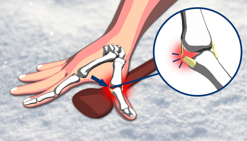
A Stener lesion is a specific and surgically important injury to the thumb. It occurs when the ulnar collateral ligament (UCL) tears completely and is then displaced by overlying tissue, preventing the ligament from reattaching to bone. Without surgical repair, a Stener lesion will not heal — making early and accurate diagnosis essential. Understanding what this injury is, why it differs from a simple UCL sprain, and how it is treated guides the entire management pathway.
What Is the Ulnar Collateral Ligament?
The ulnar collateral ligament runs along the inner (ulnar) side of the thumb’s metacarpophalangeal (MCP) joint — the knuckle at the base of the thumb. Its primary role is to stabilise the joint during pinching, gripping, and lateral loading. Without an intact UCL, the thumb deviates away from the hand under force, making the precision grip unreliable and painful.
Overlying the UCL sits a flat sheet of tendinous tissue called the adductor aponeurosis — the expanded attachment of the adductor pollicis muscle. In normal anatomy, the UCL lies deep to this aponeurosis. This anatomical relationship is the key to understanding what makes a Stener lesion distinct from other UCL injuries.
What Makes a Stener Lesion Different?

When the UCL tears partially, the ligament fibres remain in their correct anatomical position. Immobilisation in a splint allows the torn fibres to scar down and heal reliably.
When the UCL tears completely, a different problem arises. As the thumb is forced into radial deviation, the torn distal end of the UCL flips proximally, folding back on itself. The adductor aponeurosis then slides distally and interposes between the torn ligament end and its bony attachment on the proximal phalanx. The ligament is now sandwiched outside the aponeurosis — physically separated from the bone it needs to heal to.
This is a Stener lesion. Because the adductor aponeurosis acts as a mechanical barrier, the ligament cannot reattach regardless of how long the thumb is immobilised. Surgical removal of the interposed tissue and reattachment of the ligament is the only reliable treatment.
Stener lesions occur in approximately 60–80% of complete UCL tears. Not every complete tear results in a Stener lesion, which is why imaging is essential before committing to a treatment plan.
Causes of a Stener Lesion
The injury mechanism is sudden radial deviation of the thumb under load — typically a fall onto an outstretched hand, or a fall in which the thumb is caught on a ski pole as the hand hits the snow. This is why the injury is also known as skier’s thumb. The same mechanism occurs in other sports involving falls or ball contact, as well as in everyday falls onto hard surfaces.
Symptoms
Pain and swelling develop immediately at the ulnar side of the thumb MCP joint. Once acute swelling settles, patients report difficulty with pinch grip — picking up small objects, turning a key, or writing becomes uncomfortable and mechanically unreliable. Some describe a sense of instability or catching when the thumb snaags on clothing.
A small, tender, palpable nodule at the ulnar aspect of the MCP joint may be felt on examination. This represents the folded, displaced ligament lying superficial to the adductor aponeurosis — its presence strongly suggests a Stener lesion.
Diagnosis
Diagnosis begins with a history of the mechanism of injury and a focused physical examination. Stress testing applies a valgus (radial deviation) force to the thumb MCP joint in both full extension and 30° of flexion. Laxity of more than 30–35° compared to the uninjured side, or absence of a firm endpoint, indicates complete rupture. A local anaesthetic block may be needed if pain prevents adequate assessment.
The palpable nodule of displaced ligament, when present, is highly specific for a Stener lesion and an important clinical finding not to miss.
Imaging
Imaging is essential to confirm the diagnosis, quantify displacement, and exclude an associated avulsion fracture before treatment decisions are made.
X-ray is the first step. It identifies avulsion fractures — bony fragments pulled from the proximal phalanx by the torn ligament — which influence the surgical approach. A displaced avulsion fragment is itself diagnostic of a Stener lesion.
Ultrasound is the imaging tool of choice for direct visualisation of the UCL and Stener displacement in experienced hands. The torn ligament appears as a hypoechoic, folded structure lying superficial to the adductor aponeurosis — sometimes described as a “yo-yo” sign. Dynamic stress testing under ultrasound quantifies instability in real time and reliably distinguishes partial from complete tears.
MRI provides the most detailed anatomical assessment, particularly in complex or chronic cases. It confirms the position of the torn ligament relative to the aponeurosis, identifies associated chondral damage, and is the preferred investigation when ultrasound findings are equivocal or when complete soft tissue mapping is needed for surgical planning.
Treatment of a Stener Lesion
Partial UCL Tears (No Stener Lesion)
When imaging confirms an incomplete tear with the ligament in an anatomically correct position, conservative management is appropriate. The thumb is immobilised in a thermoplastic thumb spica splint or cast for four to six weeks, allowing the damaged fibres to heal. Hand therapy aims to restore range of motion, strength, and functional grip.
Complete Tears with Stener Lesion
Surgical repair is required. The adductor aponeurosis is incised, the displaced ligament retrieved and repositioned, and the torn end reattached to the proximal phalanx using suture anchors. The aponeurosis is then repaired over the ligament.
Timing is critical. Acute repairs — performed within three to four weeks of injury — allow direct ligament-to-bone reattachment with reliable, predictable results. In chronic cases, where the torn ligament has retracted, shortened, and scarred, direct repair is often no longer possible. Ligament reconstruction using a graft from the palmaris longus tendon or another donor site is then required. Outcomes after reconstruction are generally good but less predictable than after timely acute repair.
Following surgery, the thumb is immobilised in a cast for four to six weeks. Structured hand therapy then guides return to full function. Return to contact sport or skiing typically takes three to four months from the date of surgery.
The Cost of a Missed Stener Lesion
A missed Stener lesion is one of the most common causes of chronic UCL instability of the thumb. Patients present months or years later with persistent pain, weak pinch, and progressive MCP joint arthritis from chronic instability. Reconstruction remains possible in these cases, but it is technically more demanding and yields less predictable outcomes. Early diagnosis and appropriate surgical referral remain the most important factors in achieving a good result.
Final Word from Sport Doctor London on Stener Lesion
A Stener lesion is not simply a severe thumb sprain — it is a structurally distinct injury that cannot heal without surgery. The priorities are recognising it early, obtaining targeted imaging (ultrasound first, MRI if needed), and referring promptly for surgical repair before the window for direct ligament reattachment closes. Any athlete who sustains a thumb injury involving radial deviation — particularly in a fall — should have the UCL formally assessed rather than assuming it is a minor sprain.
To book a thumb assessment with Dr Lorenzo Masci at one of our central London clinics, call +44 (0) 203 488 0350 or visit our contact page. Chelsea is our most cost-effective self-pay location. Full clinic details are at sportdoctorlondon.com/clinic-locations/.
Leave A Comment