
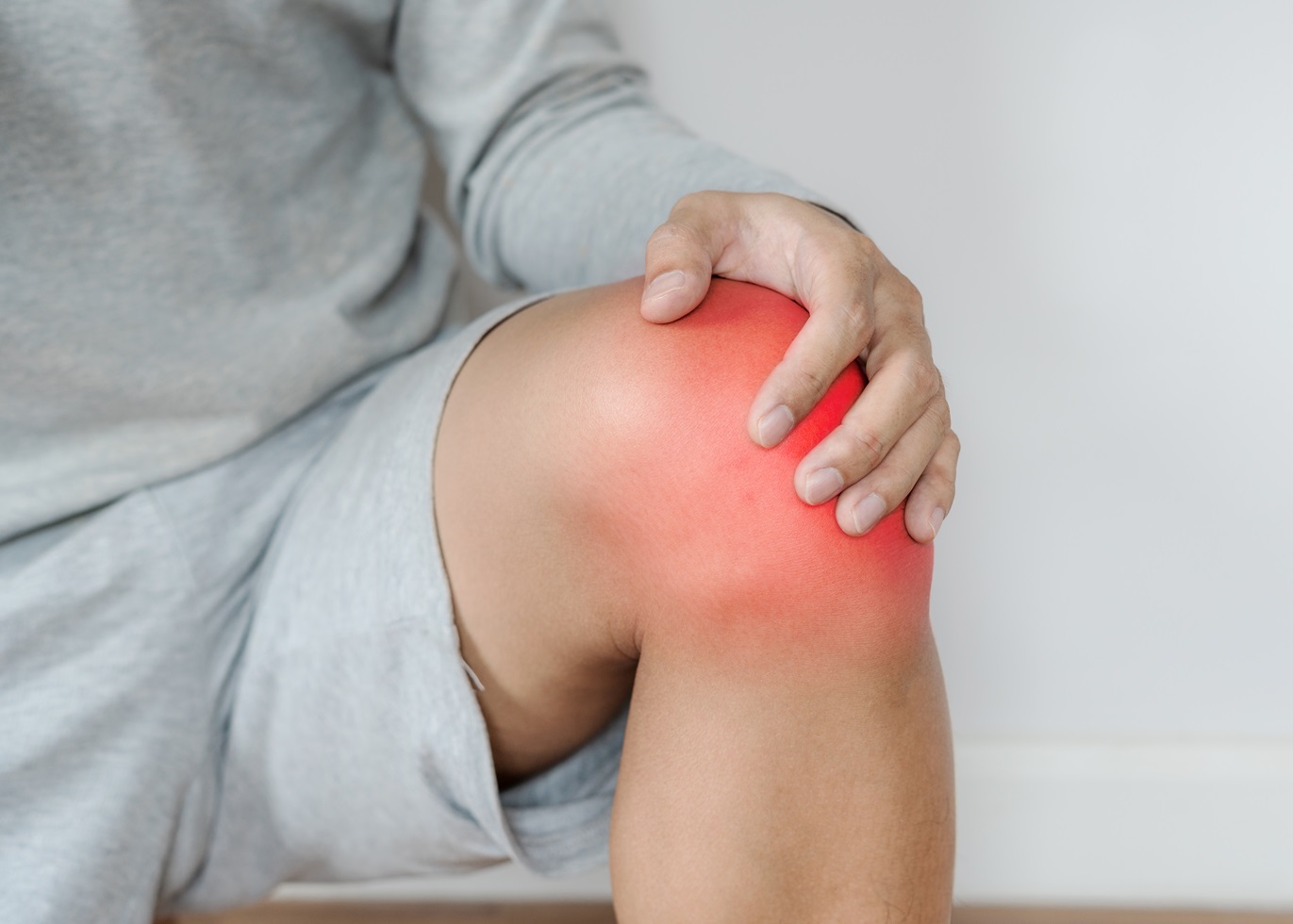
Joint pain and swelling are common in active people, usually due to cartilage damage or osteoarthritis. Occasionally, though, they come from a growth in the synovial lining of the joint, especially the knee joint. One such condition is synovial chondromatosis. So what is it, and how do we diagnose and treat it?
What is synovial chondromatosis?
Synovial chondromatosis is a rare joint condition in which small cartilage nodules form within the synovium (joint lining). Over time, these nodules can calcify and detach, creating loose bodies floating in the joint. This causes pain, swelling, and restricted movement — particularly in athletes and active people who repeatedly load their joints.
Which joints does synovial chondromatosis affect?
The knee is the most commonly affected joint, but it can also occur in the hip, elbow, shoulder, and ankle. It isn’t cancerous, but it can significantly affect joint function and, left untreated, may lead to arthritis.
Causes and risk factors
The exact cause is unknown, but it’s thought to result from abnormal growth of the synovial lining, producing excess cartilage that breaks off into the joint. Factors that may raise the risk include chronic joint stress or repetitive sporting trauma, a previous joint injury or surgery that alters the synovial environment, and inflammatory joint diseases such as rheumatoid arthritis.
There are two main types:
- Primary synovial chondromatosis — occurs spontaneously, with no underlying joint problem.
- Secondary synovial chondromatosis — develops from a pre-existing joint condition, such as osteoarthritis, trauma, inflammatory arthritis, or a joint infection.
Symptoms of synovial chondromatosis
People with synovial chondromatosis may have:
- Joint pain that worsens with activity and eases with rest.
- Swelling and stiffness from inflammation and the build-up of loose bodies.
- Clicking, locking, or catching, particularly when moving the joint.
- Restricted movement.
As they progress, calcified nodules can damage cartilage, potentially leading to early arthritis and ongoing joint problems.
How do we diagnose synovial chondromatosis?
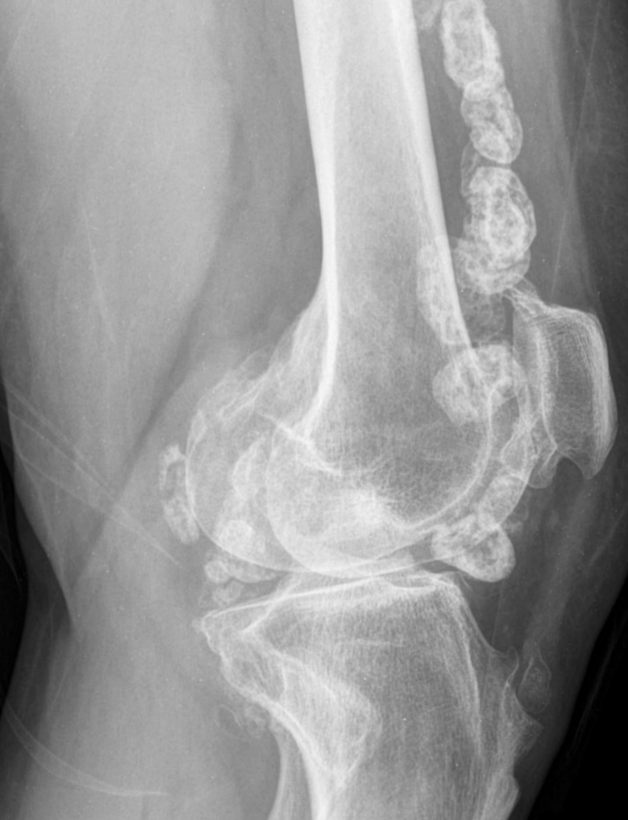
Diagnosis combines a clinical examination with imaging. Your doctor assesses the joint’s range of movement, tenderness, and stability.
Imaging confirms the diagnosis and excludes other causes. X-rays can show multiple calcified loose bodies, but may miss early cases — studies suggest 5–30% aren’t visible on X-ray. MRI is usually the most helpful, showing cartilage nodules before they calcify, assessing joint damage, and ruling out other causes. CT can also show both calcified and uncalcified cartilage bodies.
Conditions that mimic synovial chondromatosis in the knee
Because synovial chondromatosis in the knee shares symptoms with several other conditions, it’s important to distinguish it from:
- Osteoarthritis — a degenerative condition causing joint pain, swelling, and cartilage damage, but typically without loose bodies in the early stages.
- Pigmented villonodular synovitis (PVNS) — a rare synovial overgrowth that typically causes a blood-stained joint effusion.
- Inflammatory arthritis — rheumatoid or gouty arthritis, causing joint pain and swelling.
- Osteochondritis dissecans (OCD) — where bone and cartilage separate from the joint surface, sometimes forming loose bodies.
Treatment of synovial chondromatosis
Treatment depends on the severity of symptoms and the degree of joint involvement.
Non-surgical treatment
For minimal symptoms, conservative management may be enough: activity modification to reduce high-impact loading; physiotherapy to strengthen the muscles around the joint; anti-inflammatories such as ibuprofen to control pain and inflammation; and, occasionally, a corticosteroid injection for temporary relief. However, conservative treatment doesn’t remove the loose bodies so that symptoms can persist or worsen.
Surgical treatment
For severe pain, restricted movement, or locking, surgery removes the loose bodies and inflamed synovium. Keyhole (arthroscopic) surgery is suitable for localised disease; more extensive disease may require open surgery to remove all abnormal tissue. In severe cases with joint damage, joint replacement may be needed.
Can synovial chondromatosis recur?
Yes, though recurrence is uncommon, particularly in primary disease. Removing the affected synovium completely at surgery significantly lowers the risk. Continuing strengthening and mobility exercises afterwards helps maintain joint function and prevent stiffness.
Frequently asked questions about synovial chondromatosis.
How rare is synovial chondromatosis?
It affects roughly 1.8 people per million, more often men, and is most common between the ages of 30 and 50.
What is secondary synovial chondromatosis?
It’s when loose, calcified bodies form due to an underlying joint disease, such as osteoarthritis or infection. There are usually fewer loose bodies than in primary disease.
Can synovial chondromatosis become cancerous?
Very rarely. The risk is low, but malignant change should be considered in recurrent or rapidly growing lesions, which is why follow-up matters.
What’s the difference between synovial chondromatosis and loose bodies from arthritis?
Both can cause loose bodies in the joint, but synovial chondromatosis produces multiple cartilage nodules from the joint lining itself (often numerous and similar in size). In contrast, arthritis-related loose bodies arise from worn cartilage and bone. MRI and the pattern of the bodies help tell them apart.
Can I keep exercising with synovial chondromatosis?
Often, with modification, it is possible to reduce high-impact loading that aggravates the joint. But because loose bodies can cause locking and progressive cartilage damage, it’s worth getting assessed rather than training through worsening symptoms.
Final word from Sport Doctor London about synovial chondromatosis
Synovial chondromatosis is a rare condition that can limit athletic performance and daily activity. Early diagnosis with X-ray and MRI, and appropriate treatment, are key to preventing long-term joint damage. If you have unexplained joint pain, swelling, or locking, see a sports medicine doctor for assessment.
To book a joint assessment with Dr Masci in London, contact the team here or call +44 (0) 203 488 0350.
Leave A Comment