
Morel-Lavallee Lesion: How Do We Manage It?
A Morel-Lavallee lesion is a common fluid swelling that develops after trauma, with the outer hip and thigh being the usual sites. So, how do we manage a Morel-Lavallee lesion, and do you need it drained? Dr Masci diagnoses and treats these lesions at his London clinics using ultrasound-guided assessment and aspiration. This guide explains what the lesion is, how we diagnose it, and the full range of treatment options.
What is a Morel-Lavallee lesion?

A Morel-Lavallee lesion is a swelling between the underside of the skin (the dermis) and the deeper fascia. Trauma or a fall produces a shearing force across the soft tissue, tearing the blood and lymph vessels and allowing fluid to collect between the fat under the skin and the fascia. Left untreated, this fluid becomes more complex over time as the body tries to clear it.
How do we diagnose a Morel-Lavallee lesion?


A Morel-Lavallee lesion appears after trauma — classically a fall from a bicycle — where a shearing force hits the area, making contact with the ground. The lesion can appear immediately or over a few days. Other injuries sometimes occur alongside it, including fractures of the pelvis or hip, though not always.
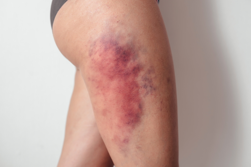
Typically, we see swelling, bruising, and skin laxity. As the fluid collection grows, it can become firm and painful. Occasionally,y it becomes infected, producing redness and heat.
The most common sites are the outer hip and the upper thigh. Lesions also appear in the lower back, the front of the knee, and over the shoulder blade.
Imagining a Morel-Lavallee lesion
Clinical examination confirms most cases, but imaging reveals fluid pockets located above the fascia. Ultrasound or MRI clearly shows these pockets, dates the lesion as acute or chronic, and rules out other injuries, such as fractures or muscle and tendon tears.
Morel-Lavallee lesion vs haematoma
Not all hip or thigh swelling is due to a Morel-Lavallée lesion. A muscle haematoma can look similar, with swelling and bruising — but its swelling sits deep within the muscle rather than above it. The clearest distinguishing sign is skin laxity, which is present in a Morel-Lavallee lesion and absent in a muscle haematoma. Other causes of hip or thigh swelling include fat necrosis, early myositis ossificans, and, rarely, a tumour.
Complications of a Morel-Lavallee lesion
Most lesions settle without problems. In some cases, though, treatment doesn’t run smoothly — complications include repeated swelling, infection, significant scar tissue, and any co-existing injuries.
Morel-Lavallee lesion treatment
Treatment depends on the size of the lesion.
Small lesions usually settle with ice and compression alone.
Larger lesions should be aspirated to remove fluid and reduce the risk of infection. We combine aspiration with ice and compression to prevent the lesion from returning — a recent study found that repeated aspiration plus compression, ice, and physiotherapy resolved most cases. Dr Masci performs aspiration under ultrasound guidance to completely remove the fluid, maintaining strict sterility to prevent infection. Aspirating more than 50 ml is associated with a higher recurrence rate.
Sclerosant injection. For larger-volume lesions (50 ml or more), we sometimes inject a sclerosant — typically high-concentration dextrose (sugar) — to encourage scar tissue to form and bind the layers together, reducing recurrence. Some practitioners use tetracycline or doxycycline instead. An honest note on the evidence: no strong trial data prove sclerosant injection beats drainage alone. In Dr Masci’s own practice, he tends to use a sclerosant alongside aspiration for larger lesions — the risk is very low, and given the high recurrence rate of big lesions, the potential benefit is worth it. It’s a pragmatic judgement rather than a purely evidence-based one.
Surgery is occasionally considered for extensive, high-volume lesions (greater than 80 ml), lesions that form a capsule, or lesions tied to other injuries.
Frequently asked questions about Morel-Lavallee lesions.
Do you need to drain a Morel-Lavallee lesion?
Not always. Small lesions resolve with ice and compression. Larger lesions — those causing a visible cosmetic bulge or growing firm and painful — should be drained, because without drainage the fluid resorbs slowly and can take many months to clear.
What is the recurrence rate after drainage?
The data vary widely, with reported recurrence anywhere from 0–75%. The key driver is volume: aspirating more than 50 ml carries a much higher recurrence rate than smaller volumes. That’s why we add compression, and often a sclerosant, for larger lesions.
Do small Morel-Lavallee lesions heal completely?
Usually, yes. Most small lesions heal fully with simple treatment, and complications such as chronic pain, infection, or scar tissue are uncommon. Persistent swelling responds well to aspiration and compression.
Is a Morel-Lavallee lesion painful?
Mild to moderate pain is normal at first from the direct trauma, settling over days to a few weeks. Severe or ongoing pain after 3–4 weeks is unusual for a Morel-Lavallée lesion and prompts us to consider another cause, including infection, which must be excluded. Long-standing lesions can ache from chronic inflammation; once infection is ruled out, a low-dose cortisone injection into the interface can help these.
How long does a Morel-Lavallee lesion take to resolve?
It depends on size and treatment. Small lesions settle within weeks. Large lesions left undrained can take many months to resorb, which is precisely why we aspirate the bigger ones rather than wait.
Can you return to sport with a Morel-Lavallee lesion?
Once the swelling settles and any larger collection has been drained, a graded return to activity is usually straightforward. Compression garments help during the return, and we confirm the lesion has not re-accumulated before full-contact or high-impact sport.
Final word from Sport Doctor London about Morel-Lavallee lesions
A Morel-Lavallee lesion forms after a shearing force or trauma to the hip or thigh. Most cases after sport or minor trauma involve small fluid volumes and settle with ice, compression, and — where needed — repeated aspiration. Larger aspiration volumes mean a higher recurrence rate, at which point we add a sclerosant or, rarely, consider surgery.
To book a one-stop assessment and ultrasound-guided aspiration with Dr Masci in London, contact his team here or call +44 (0) 203 488 0350.
My husband has this injury from a 4wheeler accident. He had multiple wash out procedures, but nothing helped and he developed an infection. It took a sclerosing procedure using doxycycline to help him. It has been 3 weeks since his most recent surgery, and it appears as though it is finally starting to heal. For reference, the accident occurred August 11, 2024 and it is now November 30th
thanks Mel for your comment. yes we use sclerosants such as high concentration dextrose but tetracycline or doxycycline (a type of antibiotic) can also be used. And yes we need to consider infection in some cases particularly for large swelling or increasing pain.
Lorenzo
I developed this recently after a traumatic accident. They measured a 3cm morel lavallee lesion along with a 10cm hematoma. I am using RICE and thankfully healing well. A lot of information I am reading online however is a bit scary and the doctors I’ve seen are largely unfamiliar with this lesion. In your experience do cases that are healing well in the acute phase resolve completely or do you see long term complications still?
Most small ML lesion heal well with simple treatments. For persistent swelling, aspiration and compression will usually resolve the lesion. Complications such as chronic pain, infection or scar tissue are rare.
Hello – I have a large morel lavallee lesion (19cmx10cm) on my buttock after a fall from a horse at the end of April. It went a month being undiagnosed. A week ago, 250ml of fluid was aspirated, and a penrose drain placed. It is still discharging serous fluid, however output has decreased overall. They are reviewing it on Monday to potentially remove drains. No sclerosing agent was administered. I am worried it will re-fill once they remove the drain. Why would they not administer a sclerosing agent to speed up tissue adherence? I want to get back to normal activities asap and it is such a slow process.
Hi Ellie, To answer your question about the sclerosing agent — the honest answer is that there is no strong evidence that sclerosant injection produces better outcomes than drainage alone. The literature in this area is limited and what exists does not convincingly show that sclerotherapy significantly reduces recurrence rates compared to drainage with or without compression. That is likely why your treating team did not administer one.
That said, in my own practice I do tend to default to using a sclerosant alongside aspiration, particularly for larger lesions like yours. My reasoning is that the risk is very low, and given the high recurrence rate associated with large lesions, I think the potential benefit is worth it even in the absence of strong trial data. It is a pragmatic rather than evidence-based decision.
LM
I have an ML lesion on my hip from a cycling accident three and a half years ago, in which I also broke two ribs, fractured a third and punctured a lung. The lesion was considered a haematoma at the time, and much less attention paid to it considering the other injuries. In spite of an infection in the area a week later, I was advised it would resolve in time. Given the area has never returned to the pre-accident size, and swells noticeably when I am unwell or run down, I have self-diagnosed the lesion – now as a 3.5 year old lesion, is treatment still possible? Thanks in advance
Thanks for your question, and sorry to hear about the accident — that was a significant set of injuries to recover from.
What you’re describing does fit a Morel-Lavallée lesion: a closed degloving injury where the skin and fat shear away from the underlying fascia and the space fills with blood and fluid. They’re very often labelled a haematoma early on, especially when more urgent injuries (like your ribs and lung) understandably take priority. The fact that it has never settled to its pre-accident size and swells when you’re run down is typical, because a chronic lesion like this develops a fibrous capsule that stops it resolving and lets it refill.
To answer your question directly: yes, treatment is still possible at three and a half years — these lesions don’t become “untreatable” with time. But the approach changes. Simple drainage alone is unlikely to be successful at this stage, because the capsule tends to refill. I have occasionally injected a small dose of cortisone into the cavity to help settle it, though I’d be cautious in your case given the infection you had in the area. Chronic lesions sometimes need a procedure to obliterate the cavity (such as sclerotherapy) or, in some cases, surgical treatment – you need up to date imaging.
LM
Dear Dr. Masci,
I have a very large lesion and would appreciate your commenting on using alcohol as a sclerant. This may be my next step, and I am very worried about it.
On May 4th, I fell through a rotting deck board at our home injuring my left leg. While there were no fractures or cuts, it was initially diagnosed as a large hematoma. It subsequently swelled significantly. On May 21st, I underwent surgery to drain and clean the area, which removed nearly 500 ml of fluid.
Unfortunately, the swelling returned after the initial drain was removed three weeks later. Since then, a radiologist has drained it again removing another 200 ml and placed a pigtail drain. Subsequent to that, he performed two rounds of sclerotherapy with doxycycline. I am returning to the radiologist this Monday to discuss removing the drain after the most recent 10 ml of doxycycline 4 days ago. I’m now draining significantly less- around 12 ml today.
Fortunately, I have not had any infections.
In two days, my doctor is going to consider taking the drain out or recommending the alcohol sclerotherapy.
I am hoping and praying we can take the drain out and use compression and see what happens.
Please let me know your thoughts on using alcohol.
Thank you!
Dear Tracey,
Thank you for reaching out and for setting out your history so clearly. I am sorry to hear what a difficult few months this has been.
To answer your question directly: alcohol is a recognised sclerosant option for lesions like yours and it is reasonable for your radiologist to consider it. That said, my own preference in these cases is to use a high-concentration dextrose (glucose) solution as the sclerosant. It works by irritating the tissue to encourage the cavity walls to adhere together, and in my experience it is effective while being associated with less local tissue toxicity than alcohol.
The fact that your drainage output has reduced to around 12ml is an encouraging sign. If the drain can be removed and the lesion does not re-expand significantly with compression, that would obviously be the preferred outcome without needing further sclerotherapy at all.
Dear Dr Masci,
I had a MML on my inner thigh following a bike accident on 7th June. It took a week to be diagnosed. Since then I have had 3* aspirations (roughly weekly) each removing 400ml of fluid. I was then advised to wear compression tights 24/7 and had weekly guided aspirations of 50, 20, 5 mls respectively. 2 weeks on from the 5ml aspiration, the lesion is now c.20 cm long by c.1-3mm deep * c. 1-2cm wide (obviously estimated from US, so not super-accurate – but indicating it’s not got bigger)
I have a few questions: at this stage, do I still need further aspirations, or will the remaining c.5mls likely resorb? If it is likely to resorb, how many weeks does it need to be ‘stable’ for before I can stop wearing the tights. Finally, I have some firm tissue in my thigh where it has seemingly healed up (i.e. no fluid accumulation). This was the point of impact from the bike handlebars ramming into my inner thigh. It is a bit painful and impedes full movement of my leg up to to abdomen. Is there anything that can be done to address this? (physio/ US to break down what I assume is scar tissue?)
Thank you for any advice!
Thank you for the detailed description; it sounds like this has been a slow but genuinely progressive recovery, with the fluid volume dropping significantly from 400ml down to just a few mls, and the lesion itself not increasing in size.
At this stage, I’d hold off on further aspirations, I think it will settle on its own from here given the trajectory you’re describing. Continue with the compression tights until the swelling in the thigh has fully settled, and I’d be guided by your treating doctors on the exact timeline for stopping them, as they’ll be able to assess the area directly.
On the firm, painful area at the point of impact, I suspect this represents significant bruising and associated tissue reaction from the direct handlebar trauma. Therapeutic ultrasound would help soften and break down this tissue, and the compression tights should also help this settle over time. Physio input would also be worthwhile alongside this, to work on soft tissue mobilisation and gradually restore full range of movement in the leg. Gentle Massage and stretching once it’s less acute can also help with this type of firm, scarred tissue.
Overall, you’re on the mend.
Dear Dr Masci
My husband has a ML on his hip after a mountain biking accident. Took 12 days to finally get diagnosed and aspirated post injury with 400ml drained. We are now 3 weeks post aspiration (4 1/2 weeks post accident) the size remained significantly smaller although still a small swelling when comparing to the other hip. NHS not offering further aspiration and the MRI but wait is 10+ weeks. My question is would you at this point pay private for an MRI and further early intervention or sit and wait it out? Im conscious the window before a capsule forming is getting smaller and smaller.
Thank you for the detailed history; this could genuinely go either way at this stage. I assume you had an initial MRI scan to confirm the diagnosis.
One option is to wait and see, given the size has already reduced significantly since the initial aspiration. The other is to proceed with an ultrasound and repeat aspiration now, and this time I’d add glucose as a sclerosant at the time of aspiration to help reduce the risk of recurrence, which addresses your concern about the window before a capsule forms.
Given the residual swelling and the long NHS wait for MRI, it may be worth considering a private ultrasound-guided assessment and aspiration sooner rather than waiting, particularly if you’re keen to act before a capsule has a chance to develop.
Dear Dr Masci,
I have had a serious MLL, due to a skiing injury (falling onto rocks) last Jan. I had it evacuated on March 20th and have gradually been recovering and making progress. I have done gentle exercises over time but upped my running to 15k a week (3x5k) in the last 3 weeks and notice the swelling is increasing and bruising appears. It seems to settle again, if I rest. Not sure if to push on or look into a further operation privately via healthcare (original was NHS). Any advice greatly appreciated. Many thanks. Nick
Thank you for the detailed history, that sounds like a significant injury to have recovered from.
Given the swelling and bruising are recurring with increased running load, and settling again with rest, I think it would be worth getting an ultrasound to look at the size of the lesion before deciding on the next course of action, which may be a repeat aspiration if there’s a significant fluid collection present.
Hi Dr Masci, I’d really appreciate your advice on a persistent thigh injury.
I fell down the stairs on 7/7 and developed a large swelling/haematoma, which was drained on 28/7; the amount was not measured.
The collection reappeared, so I had a second drainage on 10/8, with around 40 ml of fluid removed.
A subsequent ultrasound showed no remaining fluid collection, but the area is still swollen, tender and painful.
There is also a visible indentation/groove (“χαραγματιά”) with swelling above it, which is bothering me.
Could this be fat necrosis/scar tissue, and would you recommend an MRI or simply waiting for further healing/remodelling?
Thank you for the detailed history, sounds like a significant fall.
It depends on when you had the ultrasound after the second drainage. If this scan was done very recently, I’d suggest waiting and perhaps doing some rehab/physio in the meantime to help improve function, given the fluid has already been confirmed as resolved.
If the ultrasound was done immediately after the second drainage, and you think the lump or swelling has increased in size since then, I’d recommend having another ultrasound to reassess the area and check nothing has reaccumulated.
The indentation and visible groove could well represent fat necrosis or scar tissue formation following the trauma and repeated drainage, this is a recognised pattern.
I wouldn’t necessarily jump to an MRI scan unless there was something specific causing concern.
It’s important you’re assessed clinically face to face – it’s hard to advise properly without seeing you directly and reviewing your previous imaging myself.
Thank you for your quick replies to all of these questions above! I had a bike accident two weeks ago, where I went down very hard on my left shoulder and left hip. Most of my injuries have subsided, except the hip doesn’t seem to be getting better at least yet. There is still bruising and the fluid sac just below my hip. I am wondering what you consider to be a small vs a large sac. The sac itself isn’t painful, but there is a spot that is extremely painful if I press down toward the bone. I have been biking on my peloton without pain and yesterday ran without pain. Should I start icing the fluid? Is running bad?
Thanks in advance!
Thanks for the query; sounds like a heavy fall onto that hip.
If you can run without pain, then I’m not concerned about anything serious going on structurally; that’s reassuring. Ice would help the fluid sac and the tender spot.
If the swelling doesn’t settle over the coming days to weeks, you should see a doctor regarding getting an ultrasound scan to check whether you have a Morel-Lavallée lesion, given the mechanism of injury and the persistent fluid collection.
Aloha Doctor Masci, I fell off the ladder during a hurricane on August 14th and immediately developed a large lesion. At first thought it was a hematoma but after 9 days the bruising disappeared but the swelling got worse. My local Emergency room would no assit with drainage. My family doctor asprirated 220ml without any imaging guidance (ultrasound) 11 days post injury. She then drained another 50ml one week later (18 days post injury). I finally got an ultrasound and it shows the current dimensions to be 11.8 x 0.7 x 8.7 cm, My family doctor wanted to do a third aspiration without the assistance of ultrasound imaging and I declined. .07cm thickness seems too thin for my doctor to aspirate without the aid of imaging. Basic math tells me there is still 75-100ml of fluid in the lesion. Did I make the right call? I have asked to be referred to a surgeon who can aspirate with ultrasound guidance. That was 3 days ago and I still have not heard from anyone. I am continuing on with 24/7 compression but feeling very discouraged with the medical system in Hawaii. I’m worried about pseudocapsule formation (latest ultrasound did not show any thickening). With that much fluid still in the pocket, is it possible this could heal without a third aspiration or potential debridement surgery later on down the road? Thank you for your expertise, it’s a challenging lesion that nobody seems to understand out here.
Thank you for the detailed history, and sorry to hear about the difficulties you’ve had getting appropriate care out there.
I think you made the right call declining a further blind aspiration given how thin that 0.7cm dimension is, ultrasound guidance really is important for accuracy and safety in a collection that shallow. I think it’ almost impossible to drain fluid that shallow.
I’d suggest another aspiration, ideally under ultrasound guidance, with an injection of a sclerosant afterwards to reduce the chance of recurrence. I use 25% glucose for this, though other sclerosants such as polidocanol are also used. I’d avoid surgery at this stage, this can usually be managed with aspiration and sclerosant rather than needing debridement.
I’d speak to your doctor again.
All the best and good luck.
Doctor
Mi esposo sufrió una caída en bicicleta y le diagnosticaron morel la valle, le hicieron un drenaje por medio de cirugía y le dejaron una sonda por 6 días, le volvieron hacer otro drenaje y le retiraron la sonda, dándole de alta de la clínica, lleva 5 días en casa pero volvió a llenarse de líquido, se ve medianamente abultado el muslo. Tiene el control postoperatorio en 9 días, deberíamos esperar al control o ir por urgencias?
No le enviaron compresión ni drenaje
Lamento mucho lo del accidente de su marido.
La reacumulación de líquido en una lesión de Morel-Lavallée tras el drenaje es algo frecuente. No es una urgencia, salvo que su marido presente signos de infección: enrojecimiento, calor local, aumento del dolor o fiebre. Si apareciera alguno de ellos, deberían acudir a urgencias sin esperar.
Si no es el caso, esperaría a la revisión prevista, con vistas a realizar una nueva aspiración y colocar una compresión. Además, su médico podría valorar el uso de un agente esclerosante para reducir la probabilidad de que el líquido vuelva a acumularse.
I had a MLL about the size of my hand 3 years ago, which took many weeks to absorb. It made sleeping and any strenuous activity during that time uncomfortable. I tried hard to get treatment, but even though it was clearly much larger than 50ml, no-one would aspirate it.
After probably 6 months it resolved to the point of being largely unnoticeable, and after 3 years it is fine, but I can still feel a difference in the skin laxity where it was, presumably there is a capsule present and the skin is still not attached to the muscle.
I have friend who has recently suffered a similar (mountain bike) injury, reporting a “bag of water” in their buttock, which is very noticeable. They (as was I) have been diagnosed with a “hematoma”, can a “hematoma” actually present as a sloshing liquid cavity, my reading of the description precludes anything which would match this being an actual hematoma, it’s not over a joint (bursitis??), therefore it pretty much has to be a MLL? a hematoma isn’t going to be “sloshing” after 2 weeks??
Does aspiration actually speed recovery, with my MLL the doctors I saw were quite happy for me to suffer for months if it relieved them of any risk. Are most UK doctors reluctant to take any positive action, should my friend insist on a second opinion.
Thank you for your email. You ask many good questions. In essence, most doctors don’t know much about Morel-Lavallée lesions (MLL). The diagnosis is often made in accident and emergency, because most of these injuries follow major trauma, and they are frequently missed or labelled as a haematoma. I suggest aspiration, with or without a sclerosant injection, as outlined in my article here: [link]
I’m sorry to hear about your own experience. A collection the size of your hand that took many weeks to absorb, and made sleeping and exercise uncomfortable, is typical of an MLL. It is also frustrating that nobody would aspirate it. The skin laxity you still feel is very plausible. The lesion forms when the skin and fat are sheared off the fascia underneath. If a capsule formed, the layers may never have fully re-attached.
On your friend, a “bag of water” in the buttock after a mountain bike fall, still sloshing two weeks on, sounds much more like an MLL than a simple haematoma. A haematoma usually clots and feels firmer, whereas an MLL stays a fluid-filled cavity. Haematomas can liquefy over time, though, so I wouldn’t rely on the description alone. An ultrasound scan will usually tell the two apart, and an MRI can be added if needed. A bursa is also possible, but this would normally sit over a bony prominence, so it would be unusual elsewhere in the buttock.
Does aspiration speed recovery? In my experience it often does, particularly for larger or symptomatic collections. It is best done under ultrasound guidance with sterile technique, followed by compression. The evidence base is limited, and collections can recur. For those that do, a sclerosant injection to help the layers stick down, or surgery in more resistant cases, may be needed. The usual hesitation is infection risk and a lack of familiarity with the condition, rather than any proven harm from treating it.
I wouldn’t say all UK doctors are reluctant to act. Many simply haven’t seen enough of these lesions to be comfortable, and the default is to wait and see. If your friend is struggling, I would absolutely suggest a second opinion from a doctor who can do ultrasound and has experience of MLL.
LM