

If you’ve been told you have hypermobile joints, or you have widespread joint pain that no one has fully explained, Ehlers-Danlos syndrome may be worth considering. EDS is a group of inherited connective tissue disorders that remains significantly underdiagnosed — especially in sports medicine — and a missed or delayed diagnosis can have real consequences. Understanding what EDS is, how it presents, and how it’s managed is an important part of the picture for anyone dealing with persistent or complex joint pain.
What is Ehlers-Danlos syndrome?
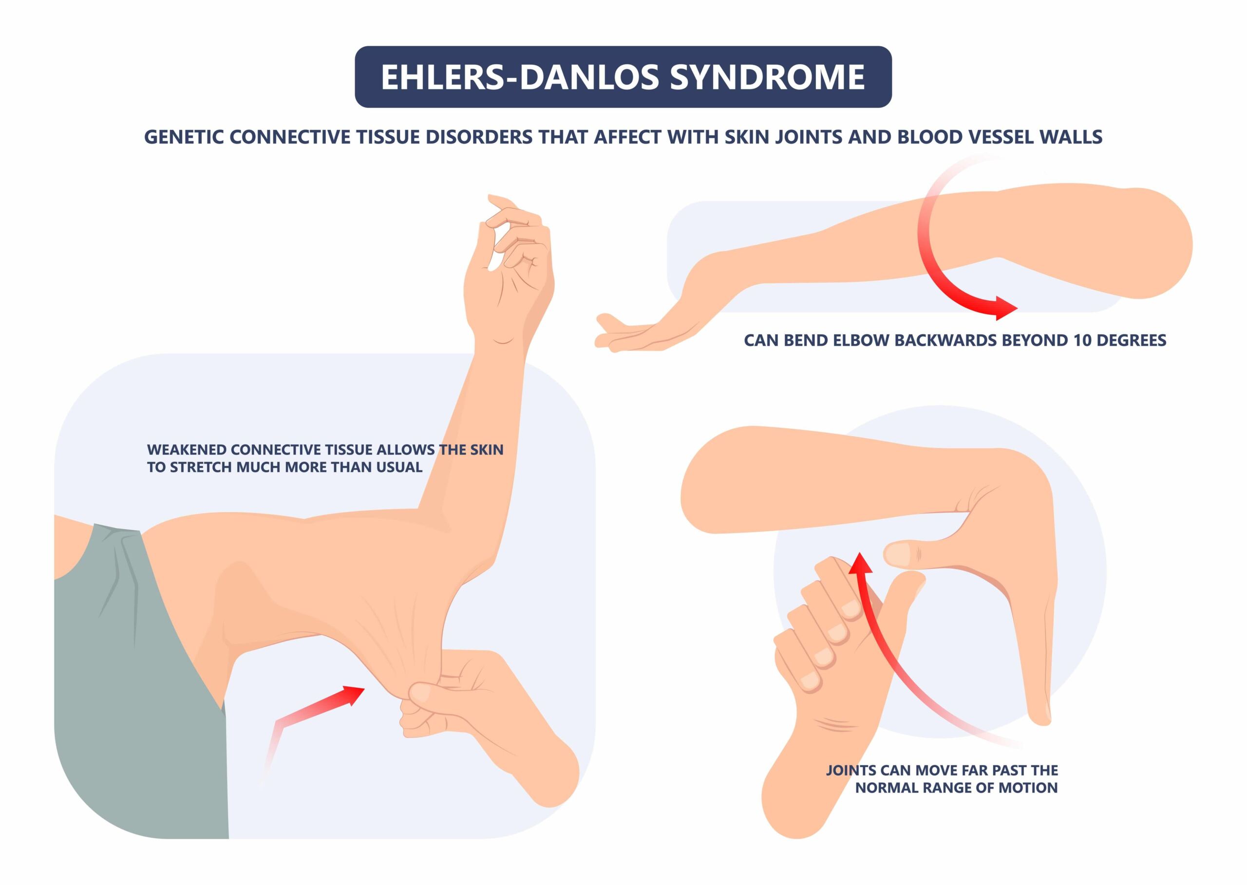
EDS isn’t a single condition but a varied group of inherited disorders caused by abnormal collagen formation. Collagen is the structural protein found throughout the body’s connective tissues — so when it’s abnormal, the effects are felt across multiple systems.
The classification recognises six main types: classical, hypermobility, vascular, kyphoscoliosis, arthrochalasia, and dermatosparaxis. The classical and hypermobility types together account for more than 90% of cases, and the hypermobility type is the most commonly seen in sports settings.
The incidence of EDS is estimated at one in 5,000 people — though this is regarded as an underestimate, given how often the condition goes unrecognised. Joint hypermobility syndrome — which many authorities now consider the same entity as hypermobility-type EDS — affects around 3% of the population.
Ehlers-Danlos and joint pain: why the link matters
Joint pain is the defining feature of EDS, and it’s particularly prominent in the hypermobility type. Chronic pain is common across all subtypes, occurring in up to 90% of patients. Moreover, pain in the hypermobility type is both more common and more severe than in the other types. It most often affects the neck, shoulders, hips, forearms, and legs — a pattern of diffuse pain rather than isolated joint disease. Most patients describe the pain as chronic (lasting more than a year) and progressive over time.
What makes EDS and joint pain so challenging is the nature of the pain. Three mechanisms operate at once:
- Mechanical pain from joint instability and dislocation, secondary to abnormal collagen.
- Soft-tissue pain from repeated micro-injury to the tendons, ligaments, and muscles around hypermobile joints.
- Nerve pain from irritation or injury of peripheral nerves, increasingly recognised as a significant contributor in the hypermobility type. This often responds better to drugs such as duloxetine or amitriptyline than to conventional anti-inflammatories.
This different origin explains why pain in EDS can be so resistant to standard approaches. No single drug or physical treatment reliably resolves it, and patients often find that several strategies each give partial relief.
The three clinical phases of hypermobility-type EDS
Hypermobility-type EDS tends to follow a course across life:
- The hypermobility phase begins early, with marked ligament laxity — often noticed in childhood or adolescence as unusually flexible joints or frequent sprains.
- The pain phase typically emerges in the second decade. Hypermobility reduces somewhat as the body stiffens protectively, while significant joint, muscle, and back pain develops and progressively limits daily activities.
- The stiffness phase develops later in life, as progressive loss of joint movement becomes the dominant feature.
This course explains why patients often reach a sports medicine clinic in the pain phase, making the diagnosis easier to miss.
How does EDS present in the sports medicine clinic?
The sports medicine clinician is well placed to spot EDS, particularly in younger patients with recurrent soft-tissue injuries. Several patterns should raise suspicion:
- Recurrent ankle sprains with minimal or no clear mechanism, or sprains that don’t settle in the expected time.
- Multiple joint injuries across different regions — shoulder instability, patellar subluxation, recurrent knee injuries — without significant trauma.
- Bilateral or multifocal symptoms in a young athlete that don’t fit a single overuse or traumatic injury.
- Poor surgical outcomes — recurrent instability after ligament reconstruction and wound-healing complications- are well documented in EDS.
- Childhood-onset pain in the shoulders, knees, ankles, or spine, which the patient may have dismissed as growing pains.
- Associated non-musculoskeletal features — easy bruising, soft or velvety skin, stretch marks out of proportion to weight change, IBS, orthostatic dizziness, headaches, and fatigue —are all recognised comorbidities of the hypermobility type.
Diagnosing EDS: the Beighton score
There’s no blood test, genetic test, or scan that confirms the hypermobility type of EDS. The diagnosis is clinical.
The Beighton score is the standard test for confirming generalised joint hypermobility. It assesses five movements, scoring up to nine points:
- Passive apposition of the thumb to the flexor forearm (one point each side)
- Passive dorsiflexion of the little finger beyond 90° (one point each side)
- Hyperextension of the elbow beyond 10° (one point each side)
- Hyperextension of the knee beyond 10° (one point each side)
- Forward flexion of the trunk, knees straight, palms flat on the floor (one point)
A score of four or more defines generalised joint hypermobility. The Beighton score falls within the broader Brighton criteria for joint hypermobility syndrome, which include major and minor criteria, such as the duration and distribution of joint pain, soft-tissue lesions, skin changes, and family history.
The score needs interpreting in context. Range of movement decreases with age, so older patients who were hypermobile in youth may score lower than their history suggests. A simple five-question screen — whether you consider yourself double-jointed, can (or could) place your palms flat on the floor, can bend your thumb to your forearm, could contort into unusual positions as a child, and whether any joint has ever dislocated — has a sensitivity of 84% and specificity of 85% for hypermobility syndrome.
Other important considerations in Ehlers-Danlos and joint pain
Several features of EDS have direct clinical implications.
Surgery carries an increased risk.
Outcomes are less good than in the general population. Pain-relieving procedures succeed in about 50% of cases, shoulder stabilisation in around 47%, and knee stabilisation in 54%. Wound-healing complications occur in about 11% of patients, and the post-operative infection rate may be up to six times higher than normal. This doesn’t mean surgery should never be done — but the threshold for operating should be higher.
Local anaesthetics may be less effective.
There’s evidence that local anaesthetics are less effective in joint hypermobility syndrome, with practical implications for diagnostic and therapeutic injection procedures.
Vascular-type EDS is life-threatening
Although the hypermobility type dominates in MSK practice, it’s vital to recognise features that might suggest the vascular type — thin skin, extensive bruising, a history of bowel or blood vessel tear, or a family history of sudden death. The vascular type has a mean life expectancy of 48 years, with spontaneous arterial rupture peaking between 30 and 40 years. Any patient with features suggestive of vascular-type EDS should be referred urgently for genetic assessment.
Cardiovascular screening matters. Everyone diagnosed with EDS should have an echocardiogram to assess for aortic root dilatation and mitral valve prolapse, both of which are more common than in the general population.
Managing Ehlers-Danlos and joint pain
Management is multidisciplinary. No treatment corrects the underlying collagen abnormality, so the focus is on reducing pain and improving function.
Physiotherapy is the cornerstone.
The goal isn’t more flexibility but the muscular strength, coordination, and proprioception needed to stabilise hypermobile joints. People with hypermobility syndrome have impaired proprioception and reduced muscle mass, both of which raise injury risk and pain. Programmes should start with a protected range of movement and progress gradually. Pool-based exercise and cycling are often well-tolerated.
Medication targets the type of pain.
NSAIDs such as ibuprofen help with inflammatory or acute flares, but need caution given the side effects. Nerve drugs such as amitriptyline and duloxetine are often needed and frequently more effective than conventional painkillers. Opioids aren’t recommended long-term due to the risk of dependence.
Supportive measures help too.
Bracing and splinting of major joints and fingers can prevent injury and pre-empt flares. Counselling often helps with anxiety and depression. Sleep is a common, under-addressed problem: poor sleep leads to increased pain. (Living with chronic pain covers some of these strategies further.)
When to refer for EDS and joint pain
A genetics consultation should be strongly considered for anyone with a Beighton score of five or above, alongside relevant symptoms, recurrent dislocations, or any personal or family history of blood vessel or organ rupture. A genetic diagnosis allows appropriate screening for vascular complications.
Frequently asked questions about Ehlers-Danlos and joint pain
Is joint hypermobility the same as Ehlers-Danlos syndrome?
Not always. Many people are hypermobile without having EDS. But joint hypermobility syndrome is now widely regarded as the same entity as hypermobility-type EDS, and it’s the joint pain, instability, and associated features — not flexibility alone — that point towards a diagnosis.
Why does Ehlers-Danlos cause joint pain?
Three types of pain often occur together: mechanical pain from unstable, subluxing joints; soft-tissue pain from repeated micro-injury around those joints; and nerve pain. This combination is why EDS pain is often hard to treat with any single approach.
How is hypermobility-type EDS diagnosed?
Clinically, there’s no confirmatory blood test or scan for this type. Doctors use the history, examination, the Beighton score (four or more of nine indicates generalised hypermobility), and the broader Brighton criteria, while excluding other causes.
What’s the best painkiller for EDS and joint pain?
It depends on the type of pain. NSAIDs help acute inflammatory flares, while nerve-type pain often responds better to neuropathic medication such as amitriptyline, duloxetine, gabapentin, or pregabalin. The most effective single measure for most people is physiotherapy.
Can exercise help EDS joint pain, or does it make it worse?
The right exercise helps. The aim isn’t more flexibility but strength, coordination, and proprioception to stabilise the joints — starting in a protected range and progressing gradually. Pool-based exercise and cycling are often well tolerated.
When should EDS be referred to a geneticist?
When the Beighton score is five or above with relevant symptoms, when there are recurrent dislocations, or when there’s any personal or family history of arterial or bowel rupture or sudden death.
Final word from Sport Doctor London about Ehlers-Danlos and joint pain
Ehlers-Danlos syndrome is an underdiagnosed cause of joint pain, particularly in younger patients with recurrent soft-tissue injuries or widespread pain that hasn’t responded as expected. Recognising it matters — it changes how injuries, surgery, and pain are managed and allows screening for more serious complications. At Sport Doctor London, Dr Masci is experienced in assessing patients with complex joint hypermobility. If you’ve struggled with persistent joint pain or recurrent injuries that haven’t responded to standard treatment, contact the team here or call +44 (0) 203 488 0350 for an assessment.
Leave A Comment